The specialty of Radiodiagnosis (better known popularly and internationally as Radiology), born in the year 1895 from the discovery of X-rays, is one of the youngest medical specialties and, nevertheless, enjoys a decisive presence and influence in the modern medicine.
According to the WHO, 80% of medical decisions in the developed world are made with the support of radiological tests. The radiologist is a clinical physician who uses medical imaging (not only X-ray but also MRI or ultrasound) to aid in the diagnosis and treatment of the patient.
What does the work of a radiologist consist of?
The key to disease treatment, and possibly the most complex process, is making an accurate diagnosis, without which treatment is never appropriate. In the vast majority of medical specialties, radiological examinations are the basis for diagnosing diseases.
The radiologist informs the doctor treating the patient about the disease or its extension, or issues a differential diagnosis and recommends further explorations to refine it. Radiology often requires the use of various imaging techniques (mammography, ultrasound, computed tomography, magnetic resonance imaging, molecular imaging) and the selection of these techniques is the responsibility of the radiologist.
In modern medicine, doctors depend on the radiologist for more than 80% of the decisions they make. The existence of well-trained radiologists is absolutely essential for any healthcare organization to offer adequate care to its patients, and that is something that all medical professionals are well aware of.
Radiologists are key to evaluating the extent of a disease. This is particularly important in malignant tumors because the degree of extension usually determines treatment. But radiology is essential in other specialties as well. For example, to advise surgeons on the appropriate technique to use in surgical procedures, or to assess the response to different types of treatment and its outcome.
Breast Cancer
Breast cancer is the most frequent malignant tumor among the female population. Raising women’s awareness of the importance of knowing breast self-examination techniques, regular check-ups and mammography are essential to detect it in time.
The importance of early detection of breast cancer through the use of mammography and other techniques is fundamental since they change the prognosis of the disease. Early diagnosis is vital because the chances of cure depend on it, which can be 100% if detected early.
How is breast cancer diagnosed?
Diagnosis of breast cancer requires microscopic examination of a sample of suspicious breast tissue (biopsy). Biopsy, however, is only the last step in a chain of procedures whose objective is to separate breast studies into two main groups: those with some degree of suspicion of cancer and those without.
To begin with, it must be said that the diagnosis of any suspected alteration of cancer is desirable before it has given signs or symptoms. The detection of cancer by palpation, although it may be in curable stages, supposes a failure of early detection. For this reason, we must insist on the recommendation that women go to their annual gynecological review and to perform an annual mammogram from the age of 40 if there is no family history, or from the age of 35 if they exist.
The history (interrogation) followed by a physical examination or physical examination of the breast is the first step taken to identify any signs of disease.
Imaging techniques
Next, some of the following imaging techniques should be used:
- Mammography
- Breast ultrasound
- Magnetic resonance
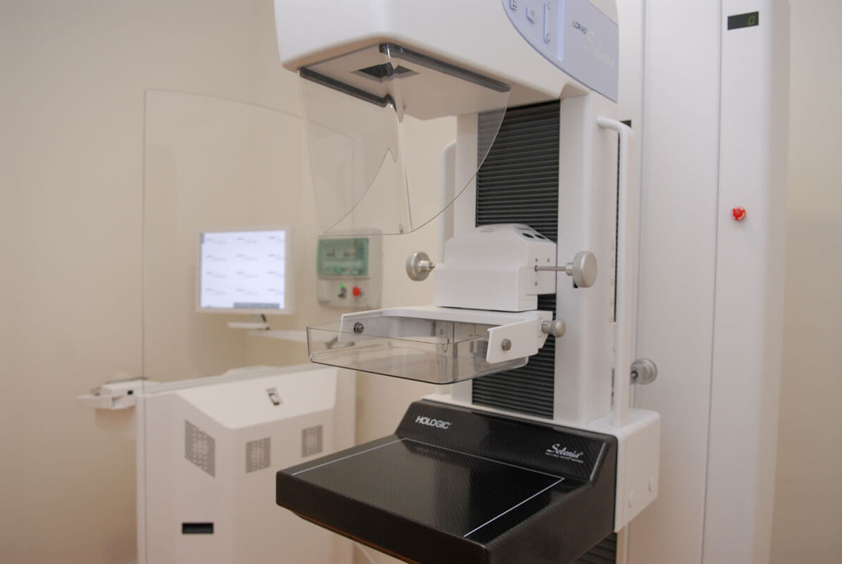
Of these procedures, the most important, most sensitive, and most widely used is mammography, an x-ray obtained on an x-ray machine that has been specially designed to study the breasts. When a mammogram is obtained, the radiologist carefully examines the images obtained looking for certain radiological signs that are known as probable indicators of pathology. The images can be obtained in an analogical way, using as a support a special radiographic film for mammography; or digitally, using digital mammography machines that directly obtain the image on digital media, which are displayed on computer screens and can be printed on film and filed on hard drives. This system allows better visualization of dense breasts and better identifies microcalcifications, one of the suspicious signs of breast cancer. It is also described as emitting less radiation than analog systems.
Breast ultrasound is a complementary method, very useful on numerous occasions, which in some circumstances may become the main diagnostic imaging technique, mainly in young women (under 30 years of age) with palpable pathology. The main utility is in distinguishing the solid or cystic nature of nodular lesions identified on mammography. It is also useful in the study of dense breast to discriminate possible injuries. It allows a very precise measurement of the size of the mammary nodules and is used to guide the punctures to obtain cellular (FNA) or tissue material (BAG) to carry out the cytological or histological examination that allows the definitive diagnosis.
Magnetic resonance imaging is important in specific cases and its use is currently not routine. However, the indications for its use are expanding more and more. The main indications for MRI are the study of multicentricity of breast cancer, assessment of local extension to support or contraindicate conservative treatment, follow-up of chemotherapy treatment, and study of complications of breast prostheses.

CENTRO DE MAMA DE TENERIFE
Centro de Mama de tenerife is the first center specialized in breast pathology in the Canary Islands. Its main objective is the early detection of breast cancer and to offer a correct diagnosis of it, carrying out the necessary procedures to achieve this end.
Our philosophy is to offer a quality service to women who come to the center so that when they leave the center they carry a normal diagnosis, or a histological diagnosis in those suspected cases, so that unnecessary time is not lost in the referral to a hospital center that performs the pertinent treatment.
Dr. Manuel Machado Calvo
Dr. Manuel Machado Calvo
Specialist in
Breast Radiodiagnosis


